The successful outcome of this project demonstrates the incredible potential bioengineering design projects from academia can have in partnership with industry.
Laurence Bray, associate chair of the Bioengineering Department

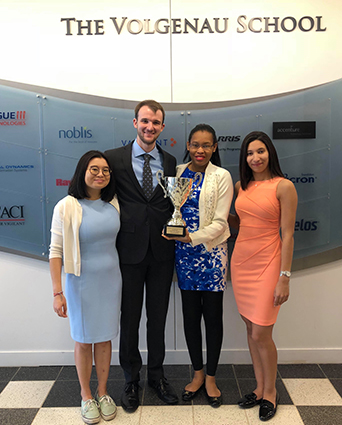
Bioengineering senior design team members Brenda Nguonly, Zachary Baker, Irvane Ngnie Kamga, and Linda Azab created an initial prototype of a system capable of merging two well-known imaging techniques. They won an innovation award for their project.
When four Mason bioengineering students launched into their senior design project last fall, they never dreamed they would build a product with the potential to improve bioengineering research, win an innovation award for their work, and rethink their career paths.
But they’ve done just that.
The team designed a device that allows one to capture both magnetic resonance imaging (MRI) and microscope images. “We’ve created an initial prototype of a system capable of merging two well-known imaging techniques, which ultimately holds implications for advancing research in the field of bioengineering,” says Zachary Baker, one of the team leaders.
The seniors won first place for their project at a recent Biomedical Engineering Society undergraduate research competition, and they plan to present their work, “Magnetic Resonance with Light Microscopy: A Dual System of Imaging,” at other conferences later this year.
Along with many other senior design groups, the team members will be presenting their poster at the Volgenau School of Engineering’s annual Undergraduate Research Celebration Monday from 5:30 p.m. to 7 p.m. at Dewberry Hall, Johnson Center. They were selected to give one of the keynote presentations at 7.
The goal of their project was to develop a novel system by modifying a compact desktop MRI system (which was invented by their sponsor Weinberg Medical Physics in Rockville Maryland) to obtain both MRI and microscope images. The MRI system is much smaller than the conventional one used for patients.
To develop the new device, the team created their own radio frequency box, integrated an inexpensive microscope into it, and inserted this in the compact MRI system. They tested the system using a bead superglued to a glass microscope slide.
The results: They were able to get an MRI image that showed the water in the bead and a microscope image that captured the details of the glue. The ultimate goal is to create a system where the images overlap one another which might one day advance biomedical research on the cellular level, Baker says.
Team leader Irvane Ngnie Kamga agrees. “Our objective was to enhance clinical research and medical diagnosis by yielding images with more features, enabling us to maximize the amount of information that can be extracted from them.”
The group headed to Rockville at 5 a.m. every Wednesday this semester to work on the device for several hours, and they were there one evening a week last semester. “We were committed. We worked so hard,” Ngnie Kamga says.
Their faculty advisor Nathalia Peixoto, an associate professor in the Bioengineering Department, says the group is the first to show proof of concept for this type of product. “It’s the beginning of an idea, but there are several significant technical challenges to be overcome.”
The successful outcome of this project demonstrates the incredible potential bioengineering design projects from academia can have in partnership with industry, says Laurence Bray, associate chair of bioengineering.
Irving Weinberg, the founder of Weinberg Medical Physics, says the team picked a challenging project and kept at it until it worked, “demonstrating the skills and attitudes that inventive engineers need in order to bring products from concepts to market.”
The students were willing to come in on nights and weekends to get the project done right, he says. "My company shares Mason’s mission to train the next generation of diverse bioengineers.”
The seniors say the experience was life-changing:
- Linda Azab was going to pursue a career in prosthetics, but she’s accepted a job at an MRI research lab at Cedars-Sinai Hospital in Los Angeles, and eventually plans to get a graduate degree in imaging. “We’re going to be telling our grandkids how our senior design project helped us figure out our careers.”
- Brenda Nguonly says she has learned the importance of persistence whether she ends up in a career in imaging or tissue engineering.
- Ngnie Kamga was intent on attending medical school in the hopes of becoming a neurosurgeon but is now considering pursuing a PhD in biomedical imaging instead.
- Baker knew he wanted to earn a PhD in bioengineering but wasn’t sure what area. He has decided to study MRI neuroimaging at Johns Hopkins University next year. “This project had a huge impact on my future.”
We’ve created an initial prototype of a system capable of merging two well-known imaging techniques, which ultimately holds implications for advancing research in the field of bioengineering.
Senior Zachary Baker
Our objective was to enhance clinical research and medical diagnosis by yielding images with more features, enabling us to maximize the amount of information that can be extracted from them.
Senior Irvane Ngnie Kamga
